What is a pulse?
EVERY time an animal’s heart beats it pushes blood out and around the body in the blood vessels.
Arteries carry blood away from the heart, delivering oxygenate blood and nutrients to the tissues, while veins transport the blood back to the heart before it travels to the lungs to collect more oxygen.
The blood in the arteries is under high pressure, as it is being pumped through them by the heart muscle. Arteries therefore have thick, muscular walls so that they don’t tear as the blood is pushed through them. This stretching of the wall of the artery each time the heart beats results in a ‘pulse’. If any artery is close to the skin we can feel this pulse by placing our fingers lightly over the artery: with each heart beat you’ll feel the artery expand under your fingertips. The number of pulses felt in a minute corresponds to the horse’s heart rate.
Early detection
There’s an additional use we can put pulse evaluation to in the horse: detecting foot problems. Injury or infection of the structures within and around the hoof is a major cause of lameness in horses, to the point where “no foot, no horse” is a well-recognised saying.
The hoof is a relatively small and highly complex structure has to be able to support the entire weight of the animal. If problems arise, the faster we can detect and begin to treat or manage them, the better the chances of recovery.
Every body part contains arteries that carry oxygenated blood to it from the heart. Those that supply the feet are called the digital arteries (‘digit’ is from the Latin term for fingers or toes). These are two digital arteries per limb – one on the inside of the leg and one on the outside. We can feel these arteries just under the skin on each leg. Because these blood vessels are a long way from the horse’s heart, the pulse in them is normally either weak or absent.
Checking the digital pulses
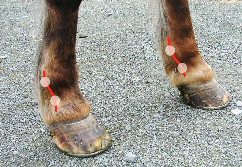
The digital arteries run down into the foot from the lower cannon region. They can be felt underneath the skin over the sesamoid bones on the back of the fetlock joint and along the pastern (pictured). To locate them, place your fingertips over the back of the fetlock joint, 2-3cm from the midline and run them from side to side over the skin.
You will feel a string-like structure under the skin. This ‘vascular bundle’ contains the digital artery, along with a vein and a nerve. Rest your fingers gently on top of it and in a normal horse you will either feel a weak pulse, or no pulse at all.
Avoid pressing firmly as this can flatten the artery and make the pulse undetectable. You can also feel for the pulses on the pastern but they may be less obvious here, especially in horses with thick skin and/or feathering.
There are two pulses per leg – one on the inside and one on the outside, giving a total of eight sites per horses that should be checked each time.
Increased digital pulses
If any tissue damage develops within the hoof the blood supply to the injured area will increase. This is part of the body’s normal healing response – damaged tissue will redden, swell, get warm and become painful.
The increased heat and redness is caused by widening of the blood vessels, allowing white blood cells to move from the circulation in to the damaged tissue and stimulate repair.
Because the tissues of the foot are enclosed by the rigid hoof wall, reddening or swelling within the foot cannot be seen. The wall may increase in temperature but this is difficult to accurately assess.
However, foot inflammation will result in a noticeable increase in the strength of the digital pulses in the affected limb. This makes digital pulse monitoring a useful way to detect foot problems early on.
Causes
Any tissue damage or inflammation in the lower limb can cause the digital pulse strength to increase. This could be as simple as a stone bruise or foot abscess, all the way up to severe laminitis, pedal bone fractures or infection of the coffin joint. In most cases, the horse will also be lame on the affected limb and prompt investigation by a vet or farrier is recommended.
Early detection of laminitis
Laminitis refers to inflammation of the laminae, the Velcro-like structures that tightly bind the pedal bone of the hoof wall. If these get inflamed to the point where they start to tear, the horse’s bodyweight will start to force the tip of the pedal bone towards the sole.
As well as being extremely painful, laminitis can be very difficult to treat, as the normal hoof blood supply and anatomy are disrupted, greatly increasing the risk of severe infection.
One of the problems with laminitis is that by the time the typical signs of the disease appear (lameness, reluctance to move, standing with the weight on the heels), the hoof has already suffered a lot of damage. However, research studies have shown that the digital pulses increase during the development phase of laminitis, 11-12 hours before any other signs are visible.
For this reason, frequent (every 3-4 hours) monitoring of the digital pulses in a horse that is at risk of developing laminitis can detect it early, and allow treatment to be started before the damage is severe.
Risk factors for laminitis include being overweight, increased weight-bearing due to lameness in the opposite leg, diarrhoea in adult horses and afterbirth that is retained for more than four hours post-foaling. Thankfully, laminitis is much less common than a foot abscess for example, but the digital pulses will be increased in any case.
By knowing how to check these pulses, owners can detect foot problems at an early stage. Practise checking the digital pulses in your horse, so that you become familiar with their normal strength and will be able to detect any abnormal changes. Pulses in the foot that are stronger than normal and persist for more than half a day may be a sign to seek treatment for the horse.
