

 This is a subscriber-only article
 It looks like you're browsing in private mode


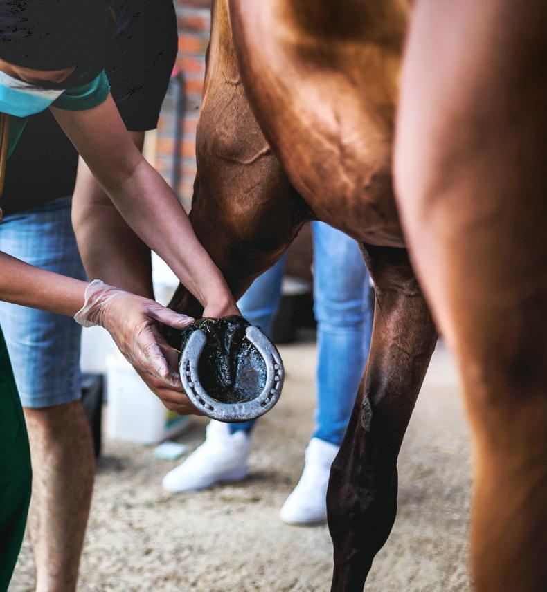
YOU’VE gone out to the paddock to bring in your horse and find them to be suddenly limping. They were absolutely fine when turned out a few hours earlier. You look at the affected leg but there’s no obvious wound, swelling or other sign of injury. You pick up the hoof; it’s cool to the touch but to your alarm, you see the head of a nail lodged in the sole. Do not pull the nail!
Everyone wants to do right by the horse, and the impulse to remove the nail immediately is completely understandable. But also potentially extremely problematic due to the risk of dirt and bacteria being carried into the vital tissues in the centre of the hoof.
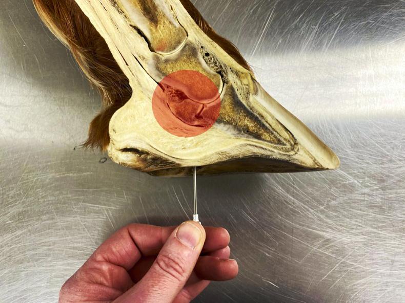
Penetrating foot injuries tend to fall into either the ‘no big deal’ or ‘absolute disaster’ categories, with very little in between. A horse’s hoof contains a lot of vital structures tightly packed into a confined space (see the area within the red shading in figure 1).
Deep flexor tendon
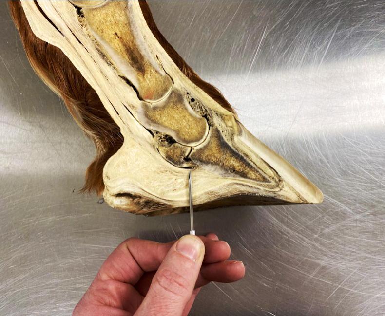
The distance between the coffin joint and the sole is only about one to two centimetres. Layered around the coffin joint are the deep flexor tendon and its enveloping tendon sheath, the navicular bone, the navicular bursa (cushioning sac), various ligaments and the enveloping ‘Velcro-like’ laminae that bind the hoof wall tightly to the coffin bone. It’s relatively easy therefore for a sharp object to pierce the sole and prick or tear one or more of these structures (figure 2).
Deep-seated foot infections, especially within the coffin joint, tendon sheath or navicular bursa are potentially both career-ending and life-threatening, as any infection-induced scarring can prevent these structures gliding smoothly over each other, resulting in chronic pain and severe lameness.
Horses need to bear weight on all four legs - they do not make a good tripod! If the affected leg is very painful it can lead to laminitis or tendon failure in the supporting limb. The welfare of a horse with only two good legs is so severely impacted that humane euthanasia may become the only viable option.
The closer any damage is to the edge of the sole, the less likely it is to be very serious. The problem is that what started out as a straight nail or piece of metal can readily buckle under the weight of the horse and turn inwards, heading for the delicate structures tucked away in the centre of the hoof.
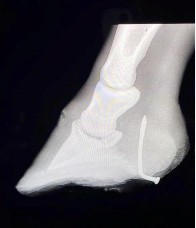
Foot X-ray
Where possible, leave the nail in place until a veterinary examination can take place. A foot X-ray with the nail in situ will quickly reveal its direction of travel, and help figure out the most appropriate course of treatment. Ideally, keep the horse from moving around and causing further damage while waiting for assistance. Clean, dry footing will help keep the wound from getting further dirt packed into it. Two rolls of bandage, or a thick piece of aeroboard with a hole cut in it, can be placed over the sole either side of the injury site and taped or bandaged in place to help prevent the horse driving the object further into its foot.
If prompt veterinary assistance is not available, you should take several photographs of the nail and sole to record its precise location and orientation, before pulling it out (a pliers may be needed). Use a hoof knife to pare away a little of the sole around the wound, so that it can be accurately located by the vet later. Clean and wrap the foot and save the nail to show how far in it went. You’ll also need to retrieve the horse’s passport or vaccination records, to allow the vet to check if the horse’s tetanus protection is adequate.
If there is any chance that the critical structures in the foot have been damaged, then the horse should be admitted to a veterinary hospital for more specialised imaging and diagnostic tests. Surgical intervention under a general anaesthetic may be required to flush the coffin joint or tendon sheath, trim away infected tissue, and allow the wound to drain.
Foot cast
A technique where a tourniquet is applied to the leg so that high concentrations of antibiotics can be instilled into the foot may also be helpful. A foot cast or sterile bandage is applied while healing occurs and a special shoe may also be needed. Recovery can take several months and not all of these horses are able to return to their previous level of performance. So a nail in the foot may indeed be why the horse was lost!
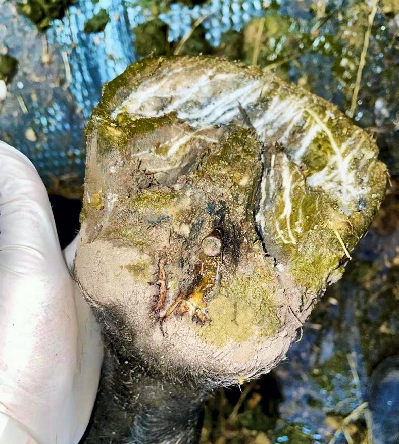
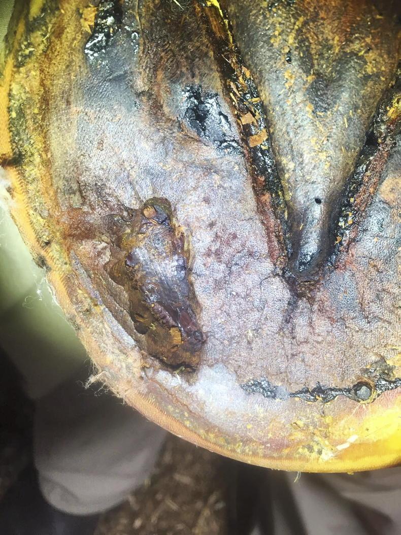
Figure 3 is a photo of a nail that was found to have pierced the base of a horse’s frog, close to the heel bulbs. The angle of entry on closer inspection looked very ominous, with the direction of travel appearing to be towards the navicular bone. The X-ray (figure 4) revealed however, that this horse had been lucky and the nail had bent away from the bone and was instead stuck in the heel bulbs – painful no doubt, but not life-threatening. The nail was removed and the tract in the hoof cleaned and flushed. The foot was dressed and bandaged and a course of antibiotic therapy, a tetanus booster, and pain relief happily resulted in a full recovery for this horse.


SHARING OPTIONS: